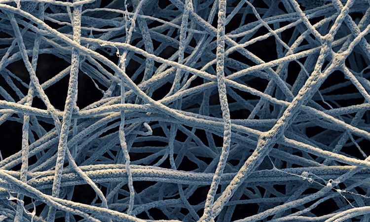
There’s a quiet corner of materials science where carbon fibres get dressed up in graphene, and the result is a biosensor that’s sharper, cheaper, and considerably more interesting than what came before it. This is that corner.
Biosensors are, at their core, translators. They take biological information — the presence of an enzyme, a virus, a hormone — and convert it into an electrical signal that a machine can read and display. The better the electrode at the heart of the biosensor, the more clearly it can “hear” what the biology is saying. Researchers at the University of Edinburgh tackled this problem not by reinventing biosensors entirely, but by rethinking what the electrode is made of.
Their approach: carbon nanofibers (CNFs), wrapped in graphene.
What Are Carbon Nanofibers, and Why Should You Care?
Carbon nanofibers are exactly what they sound like — fibres made of carbon, at the nanoscale. They have a large surface area (useful for sensing), decent electrical conductivity, and they’re chemically stable, meaning they don’t fall apart the moment something interesting happens around them. In biosensing, that’s rather important.
The team created their CNFs using a technique called electrospinning: essentially, you push a polymer solution through a fine tip using an electric charge, and it sprays out as a mat of extremely thin fibres onto a rotating plate. Think of it as a very controlled, very scientific candy-floss machine. The fibres were then baked at 200°C to stabilise them, and carbonised at 750°C in an oxygen-free environment — a process that converts the polymer backbone into actual carbon.
Enter Graphene
Here’s where it gets more interesting. Graphene — a single layer of carbon atoms arranged in a hexagonal lattice — is one of the most electrically conductive materials known. Wrapping CNFs in graphene is essentially giving them an upgrade: more surface area for reactions to occur, and significantly better conductivity.
The team did this by submerging their fibres in a graphene oxide solution, adjusting the pH and concentration, then adding a reducing agent (hydrazine monohydrate) to convert the graphene oxide into graphene — which bonded directly to the fibre surface as it did so.
The results were measurable. Under electron microscopy, the smooth fibres became visibly rougher and wrinkled after wrapping, consistent with a graphene coating. More meaningfully, their electrochemical testing showed the graphene-wrapped, carbonised electrode had over twice the electroactive surface area of a bare reference electrode, and was more than 14 times more conductive — even accounting for limitations introduced by the carbon glue used to attach the sample.
Why This Matters
Better conductivity means a biosensor can detect smaller amounts of a target molecule, more reliably. Electrospinning is also relatively inexpensive and scalable — it doesn’t require the kind of equipment that makes lab-to-clinic translation prohibitively slow or costly.
This isn’t a cure. It’s not even a product yet. It’s a proof of concept that an improved electrode material — cheap to make, better at its job — can be fabricated using accessible methods. That’s what incremental materials science looks like, and incremental is often exactly what diagnostics needs.
Sources
- Tsiamis et al. (2021). Graphene Wrapping of Electrospun Nanofibers for Enhanced Electrochemical Sensing. ACS Omega. https://pubs.acs.org/doi/10.1021/acsomega.0c05823
- Pumera, M. (2011). Graphene in biosensing. Materials Today. https://doi.org/10.1016/S1369-7021(11)70160-2
- Persano, L. et al. (2013). Industrial upscaling of electrospinning and applications of polymer nanofibers. Macromolecular Materials and Engineering. https://doi.org/10.1002/mame.201200197
- Novoselov, K.S. et al. (2004). Electric field effect in atomically thin carbon films. Science. https://doi.org/10.1126/science.1102896

